


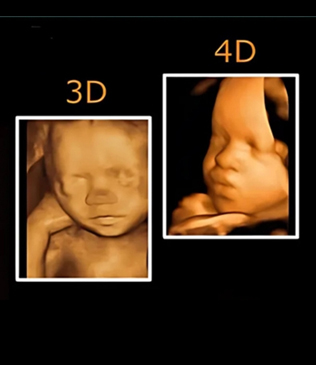
3D & 4D Ultrasound
3D and 4D ultrasound are advanced imaging techniques used during pregnancy to obtain detailed images of the developing baby inside the womb. Unlike traditional 2D ultrasound, which provides flat images, 3D ultrasound produces three-dimensional images of the baby, while 4D ultrasound provides real-time moving images. This allows doctors and parents to observe the baby's facial features, movements, and overall development more clearly.
These advanced ultrasound technologies are commonly used for both medical evaluation and enhanced visualization during pregnancy. They help healthcare professionals assess fetal development, detect certain abnormalities, and monitor the baby's growth in greater detail. In addition to medical benefits, 3D and 4D ultrasounds also allow parents to see their baby’s movements such as yawning, stretching, or smiling in real time.
Benefits of 3D & 4D Ultrasound
- Provides clearer and more detailed images of the baby
- Helps detect certain structural abnormalities
- Allows better visualization of fetal facial features
- Real-time monitoring of fetal movements
- Improves assessment of fetal development
- Enhances bonding experience for parents
What Can Be Evaluated with 3D & 4D Ultrasound
- Fetal facial structures and features
- Baby’s movements and activity
- Growth and development of the fetus
- Position of the baby in the uterus
- Placental location and condition
- Amniotic fluid levels and surrounding environment
When 3D & 4D Ultrasound is Recommended
- Detailed fetal anomaly assessment
- Monitoring fetal development during pregnancy
- Evaluating facial or structural abnormalities
- High-risk pregnancy evaluation
- Additional visualization after routine ultrasound
- Special medical recommendations by the doctor
Procedure Overview
- A gel is applied to the abdomen for better imaging
- An ultrasound probe is gently moved across the abdomen
- Sound waves create detailed images of the baby
- 3D images display the baby's structure
- 4D technology shows live movements in real time
- The procedure is safe, painless, and non-invasive
Safety and Care
- Ultrasound uses safe sound wave technology
- No radiation is involved in the procedure
- Performed by trained sonographers or medical professionals
- Recommended only when medically appropriate
- Safe for both mother and baby when performed correctly
- Regular prenatal check-ups should continue as advised
3D and 4D ultrasound technology has greatly improved prenatal imaging, providing detailed insights into fetal development and helping doctors monitor pregnancy more effectively. With advanced imaging capabilities and expert medical care, these ultrasounds support better diagnosis, improved pregnancy monitoring, and a memorable experience for expecting parents.